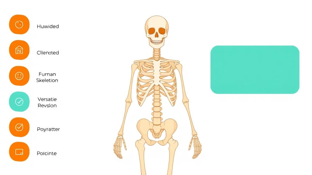
Human Skeleton Labeler turns complex biology into clear, memorable explanations your child can actually understand. It uses simple language and helpful analogies to break down every topic — from cell structure and genetics to ecosystems and the human body.
Photosynthesis is how plants make their own food! Think of a leaf as a tiny solar-powered kitchen:
1. **Ingredients:** Water (absorbed by roots) + Carbon Dioxide (absorbed from air) + Sunlight (captured by green chlorophyll in leaves).
2. **Cooking:** Sunlight energy powers a chemical reaction that combines water and carbon dioxide.
3. **Food Served:** Out comes **Glucose** (sugar energy for the plant to grow) and **Oxygen** (which they release into the air for us to breathe!).
Equation: Water + Carbon Dioxide + Light ➔ Glucose + Oxygen. Pretty amazing, right?
How to use Human Skeleton Labeler
1
Open Applaa and select this tool from AI Assistants.
2
Describe the biology topic you're studying or paste a question from your textbook.
3
Get a clear explanation with real-world examples — ask any follow-up question as many times as needed.
What you can do with Human Skeleton Labeler
Learn the names of bones. Here are some of the most popular ways students use Human Skeleton Labeler every day:
Makes complex biology genuinely accessible at every level
Covers GCSE, A-Level, and IB Biology comprehensively
Great for revision, homework help, and science fair projects
Ask unlimited follow-up questions — the AI never loses patience or gives up on you
Works alongside any textbook, worksheet, or school resource
The Human Skeleton Labeler is your interactive anatomy atlas, identifying every bone, cartilage, and major ligament in the human skeleton—from the tiny ossicles in your ear to the femur (longest bone) in your thigh. GCSE and A-Level Biology expect solid understanding of skeletal anatomy and function; many UK students struggle with the 200+ bones and their interrelationships. Rather than flipping textbook pages, Applaa's Human Skeleton Labeler lets you rotate a 3D skeleton model, zoom into specific regions (skull, spine, limbs), and click bones to reveal their names, functions, and connections to muscle and movement. You'll learn that bones aren't dead structures—they're dynamic: they store calcium and phosphorus, produce blood cells in marrow, and remodel constantly in response to stress. The labeler emphasises the skeleton's roles: support (framework for the body), protection (skull shields the brain; ribs protect organs), movement (bones lever with muscles), and mineral storage. Essential for GCSE and A-Level Biology, sports science, and medicine pathways.
206
Adult human bones with individual labels and functions
3D
Interactive model with full rotation, zoom, and regional focus
100%
Aligned to GCSE and A-Level anatomy curricula
How to use Human Skeleton Labeler effectively
Start with the full-skeleton view, rotating the model to familiarise yourself with overall structure: skull, vertebral column, ribs, pelvis, limb bones. Next, zoom into the skull and label key bones: cranium, mandible, maxilla, zygomatic (cheekbone). Move to the vertebral column: cervical (neck), thoracic (chest), lumbar (lower back), sacrum (fused vertebrae at base), coccyx (tailbone). Then explore the limbs: humerus (upper arm), radius and ulna (forearm), carpals (wrist), metacarpals (palm), phalanges (fingers). Use the 'Associated Structures' feature to see muscles attached to each bone—the biceps inserts on the radius; the deltoid inserts on the humerus. Study the joints where bones meet: show how the ball-and-socket hip allows rotation, whilst the hinge elbow allows flexion-extension. Finally, revisit regions weekly, testing yourself by hiding labels and recalling bone names and functions.
Learn bone names in anatomical position: standing upright, arms at sides, palms forward—this standardises terminology
Master the major bones first (femur, humerus, tibia, fibula, radius, ulna, carpals, tarsals) before memorising smaller ones
Understand each bone's role: femur supports body weight and transmits force from jumping; clavicle braces the shoulder; vertebrae protect the spinal cord
Link skeleton to muscle: every muscle has origin (on stable bone) and insertion (on moving bone); contraction pulls the insertion toward the origin, creating movement
Note skeletal features: condyles (rounded surfaces for joints), tuberosities (rough bumps where muscles attach), foramina (holes for nerves and blood vessels)
Study joint types: ball-and-socket (hip, shoulder) for rotation; hinge (elbow, knee) for flexion; pivot (atlas-axis, atlanto-occipital) for rotation
Common mistakes with Human Skeleton Labeler
Students often memorise bone names in isolation, missing their functions and relationships. For example, they learn 'femur' as a fact without understanding it's the longest, strongest bone because it bears half the body's weight when standing. Another trap is viewing the skeleton as static; in reality, bones remodel throughout life in response to stress, calcium intake, and hormones—why teenage athletes' bones strengthen with training, and why elderly people with low calcium develop brittle bones. UK learners sometimes confuse similar bone names (radius vs. ulna, tibia vs. fibula) without remembering which is lateral (thumb/fibula) and medial (pinky/tibia). Many also overlook bone functions beyond support: producing red blood cells in marrow, storing minerals, and maintaining blood calcium levels. Finally, some students struggle to visualise 3D relationships from 2D diagrams, missing joint angles and rotation axes.
Don't memorise in a vacuum: link each bone to its function, muscle attachments, and movement role
Recall that the human skeleton has 206 bones in adults (some fuse with age); infants have ~270 (cartilaginous), which ossify over years
Remember 'lateral' and 'medial' positioning: radius is lateral (thumb side) in anatomical position; ulna is medial (pinky side)
Understand bone remodelling: stress stimulates osteoblasts to build bone; lack of stress (immobilisation, microgravity) allows osteoclasts to dissolve it
Link skeleton health to diet and exercise: calcium and vitamin D are essential for bone formation; weight-bearing activity stimulates remodelling
Visualise joints in 3D: a hinge joint moves in one plane (elbow flexes/extends); a ball-and-socket moves in multiple planes (shoulder abducts, adducts, rotates)
Getting started
Getting started with Human Skeleton Labeler
Step 1
Download Applaa free and open 'Human Skeleton Labeler' under Biology & Life Science
Step 2
Rotate the full skeleton model to orient yourself; then focus on the skull and label cranium, mandible, maxilla, and temporal bones
Step 3
Study the vertebral column: identify cervical, thoracic, lumbar, sacral, and coccygeal regions; understand why lumbar vertebrae are largest (weight-bearing)
Step 4
Zoom into limbs and practice naming humerus, radius, ulna, carpals, phalanges (arms) and femur, tibia, fibula, tarsals, metatarsals (legs) until you recall them without hints
1 month free, then 50% off for 3 months — £4.99/mo
While Other Kids Get an AI Head Start, Is Yours Falling Behind?
Give your child the same AI app-building, exam prep and tutoring edge — free for the first month, no credit card needed.
Join 10,000+ students building their first AI-powered apps with Applaa
Frequently asked questions about Human Skeleton Labeler
Why do exam questions ask us to identify bones we could just look up?
Because understanding bone anatomy is the foundation for understanding movement, injury, and physiology. If you can't name the femur and tibia, you can't explain how a knee injury affects your walking, or how exercise strengthens leg bones. Examiners test this knowledge to ensure you grasp anatomy's relevance to health and biomechanics.
How does bone density relate to GCSE health topics?
Bone density is crucial for osteoporosis questions. Our labeler emphasises that bone is living tissue constantly remodelling. Calcium, vitamin D, and exercise strengthen bone; sedentary lifestyles and low calcium weaken it. This ties to A-Level physiology, nutrition, and public health—especially for ageing populations.
Can I use the 3D model to explain joint movement for A-Level biomechanics?
Yes. The labeler shows how bones articulate at joints and where muscles attach. Understanding that the biceps pulls the radius closer to the humerus (flexion) or that the deltoid lifts the humerus (abduction) requires visualising the 3D skeleton. Our interactive model is ideal for building this spatial understanding.
Does the labeler cover special anatomical features like the greater trochanter or olecranon?
Yes. These landmarks are where muscles attach or where bones articulate. The greater trochanter (bump on the femur) is where hip muscles attach; the olecranon (bony point of the elbow) is where the triceps inserts. Our tool labels these features and explains their significance for muscle action and exam questions about skeletal anatomy.